
|
Contact |
FAQ |
Jobs
|
|
|

|
Eye of Horus |
The multiple paths leading to the pyramid reflect on both a multidisciplinary
approach and the importance of including diversity in conducting scientific research.
The pyramid reflects on “elements” forming a greater whole. The numbers in the back
of the pyramid reflect on our love for mathematics and quantitative assessment.
Clicking on the Eye of Horus label provides insight into “Why the Eye of Horus” as
a logo for the ODALab [Courtesy graphics of Dr. Catherine Meyer, an ODALab Research
Team Member in the late 1990s]. Surely, the “Eye” also serves well the ODALab spectrum
where optics is a core area of research.
Under the Rochester tab you can find more about the lab members, and among other activities you can access
our publications.
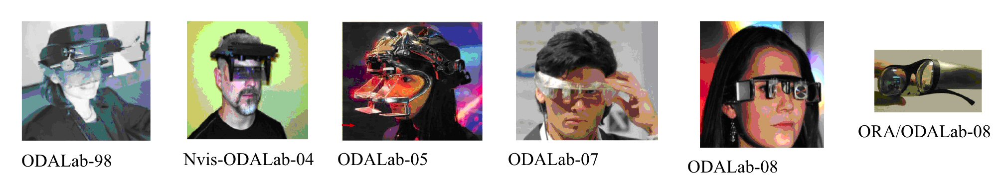
History of HWDs in ODALab
Funding Highlights (≥500K)
-
2009: NYSTAR Foundation funds the ODALab-Rochester in the area of Biotechnology [2009-2011] ($949,883)
Prof. Rolland recognized as a NYSTAR Fellow.
-
2008: The James and Esther King Biomedical Research Program funds MD Anderson Cancer Center Orlando
and the ODALab-Orlando for Medical Research related to Physics-based Modeling of Breathing Lungs and
Lung Cancer Treatment [2008-2010] ($949,717) - The Florida I4 Corridor provides matching funds for the project.
- 1997: FIRST AWARD from the NIH/NLM ($500K)
Notable Events
-
2009 The ODALab Headquarter moves to the University of Rochester.
-
2009 Satellite ODALab-Orlando opens led by Dr. Anand Santhanam.
-
2006 Satellite ODALab-Georgia (NEWS) opens at the Armstrong Atlantic University in Georgia led by Dr. Felix Hamza-Lup.
Historical Notes
In 1999, the M.I.N.D. Labs and ELF-France Acquitaine provided together the funding required for the development of our first Head-mounted Projection Display (HMPD) and the first most compact projector at the time was designed that can be worn on the head with 6g per eye lenses.
We created content for displays especially in the area of medical imaging and military training - see our MEDIA for the knee-joint video as part of the Virtual Reality Dynamic Anatomy (VRDA) tool – A project first funded by the NIH/NLM as a FIRST AWARD from 1997-2002.
The early 2000’s, the Research in the ODALab expanded to include research in optical imaging, such as in Optical Coherence Tomography (OCT). This area of research has bloomed to an active area of research in the ODALab where we focus on instrumentation development for high lateral-resolution imaging across extended depth-of-focus via Bessel Beam Imaging and Gabor Domain Optical Coherence Microscopy (GD-OCM).
The 2010’s - Recently, research in the ODALab, driven by critical needs, among others, in the applications of AR and medical research has started focused efforts in non-imaging optics, and optical design, fabrication, and testing of freeform optics.
Several national and international sponsors, collaborators and industrial partners participate in the research efforts. Governmental support is given through grants from the National Science Foundation (NSF), the National Institute of Health (NIH), the NYSTAR Foundation, STRICOM, the US Army Research Office, and the Office of Naval Research (ONR), and private Foundations. Finally many industrial partners have committed to join force with the ODA Lab.
Today, we are partnering with Medical Centers to conduct research related to the Upper Airways [MD Anderson Cancer Center Orlando], the Human Eye [URMC], Skin [URMC], and the Oral Cavity [URMC].